Diagnosis
Scoliosis diagnosis treatment is usually confirmed through a physical examination, an x-ray, spinal radiograph, CT scan, or MRI. The curve is measured by the Cobb Method and is diagnosed in terms of severity by the number of degrees. A positive diagnosis of scoliosis is made based on a coronal curvature measured on a posterior-anterior radiograph of greater than 10 degrees. In general, a curve is considered significant if it is greater than 25 to 30 degrees. Curves exceeding 45 to 50 degrees are considered severe and often require more aggressive scoliosis diagnosis treatment.
A standard exam that is sometimes used by pediatricians and in grade school screenings is called the Adam’s Forward Bend Test. During this test, the patient leans forward with his or her feet together and bends 90 degrees at the waist. From this angle, any asymmetry of the trunk or any abnormal spinal curvatures can easily be detected by the examiner. This is a simple initial screening test that can detect potential problems, but cannot determine accurately the exact type or severity of the deformity. Radiographic tests are required for an accurate and positive diagnosis.
-
- X-ray : Application of radiation to produce a film or picture of a part of the body can show the structure of the vertebrae and the outline of the joints. X-rays of the spine are obtained to search for other potential causes of pain, i.e. infections, fractures, deformities, etc.
-
- Computed tomography scan (CT or CAT scan): A diagnostic image created after a computer reads X-rays; can show the shape and size of the spinal canal, its contents and the structures around it. Very good at visualizing bony structures.
-
- Magnetic resonance imaging (MRI) : A diagnostic test that produces three-dimensional images of body structures using powerful magnets and computer technology; can show the spinal cord, nerve roots and surrounding areas, as well as enlargement, degeneration and deformities.
In Children
Scoliosis in children is classified by age: 1.) Infantile (0 to 3 years); 2.) Juvenile (3 to 10 years); and 3.) Adolescent (age 11 and older, or from onset of puberty until skeletal maturity). Idiopathic scoliosis comprises the vast majority of cases presenting during adolescence. Depending on its severity and the age of the child, scoliosis is managed by close observation, bracing and/or surgery.
In children with congenital scoliosis, there is a known increased incidence of other congenital abnormalities. These are most commonly associated with the spinal cord (20 percent), the genitourinary system (20 to 33 percent) and the heart (10 to 15 percent). It is important that evaluation of the neurological, genitourinary and cardiovascular systems is undertaken when congenital scoliosis is diagnosed.
In Adults
Scoliosis that occurs or is diagnosed in adulthood is distinctive from childhood scoliosis, since the underlying causes and goals of treatment differ in patients who have already reached skeletal maturity. Most adults with scoliosis can be divided into the following categories: 1.) Adult scoliosis patients who were surgically treated as adolescents; 2.) Adults who did not receive treatment when they were younger; and 3.) Adults with a type of scoliosis called degenerative scoliosis.
In one 20-year study, about 40 percent of adult scoliosis patients experienced a progression. Of those, 10 percent showed a very significant progression, while the other 30 percent experienced a very mild progression, usually of less than one degree per year.
Degenerative scoliosis occurs most frequently in the lumbar spine (lower back) and more commonly affects people age 65 and older. It is often accompanied by spinal stenosis, or narrowing of the spinal canal, which pinches the spinal nerves and makes it difficult for them to function normally. Back pain associated with degenerative scoliosis usually begins gradually and is linked with activity. The curvature of the spine in this form of scoliosis is often relatively minor, so surgery may only be advised when conservative methods fail to alleviate pain associated with the condition.
Treatment
When there is a confirmed scoliosis diagnosis treatment, there are several issues to assess that can help determine treatment options:
-
- Spinal maturity – is the patient’s spine still growing and changing?
-
- Degree and extent of curvature – how severe is the curve and how does it affect the patient’s lifestyle?
-
- Location of curve – according to some experts, thoracic curves are more likely to progress than curves in other regions of the spine.
-
- Possibility of curve progression – patients who have large curves prior to their adolescent growth spurts are more likely to experience curve progression.
After these variables are assessed, the following treatment options may be recommended:
-
- Observation
-
- Bracing
-
- Surgery
Observation
In many children with scoliosis, the spinal curve is mild enough to not require treatment. However, if the doctor is worried that the curve may be increasing, he or she may wish to examine the child every four to six months throughout adolescence.
In adults with scoliosis, X-rays are usually recommended once every five years, unless symptoms are getting progressively worse.
Bracing
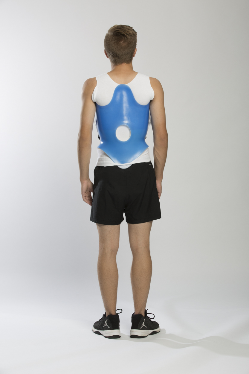
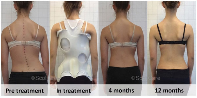
Braces are only effective in patients who have not reached skeletal maturity. When there is a confirmed scoliosis diagnosis treatment, if the child is still growing and his or her curve is between 25 degrees and 40 degrees, a brace may be recommended to prevent the curve from progressing. There have been improvements in brace design, and the newer models fit under the arm, not around the neck. There are several different types of braces available. While there is some disagreement among experts as to which type of brace is most effective, large studies indicate that braces, when used with full compliance, successfully stop curve progression in about 80 percent of children with scoliosis. For optimal effectiveness, the brace should be checked regularly to ensure a proper fit and may need to be worn 16 to 23 hours every day until growth stops. A scoliosis diagnosis treatment plan often includes bracing for younger patients with moderate curves to prevent further progression.
Surgery
In children, when there is a confirmed scoliosis diagnosis treatment, the two primary goals of surgery are to stop the curve from progressing during adulthood and to diminish spinal deformity. Most experts would recommend surgery only when the spinal curve is greater than 40 degrees and shows signs of progression. This surgery can be done using an anterior approach (through the front) or a posterior approach (through the back), depending on the particular case.
Some adults who were treated as children may need revision surgery, especially if they were treated 20 to 30 years ago, before major advances in scoliosis diagnosis treatment procedures were implemented. In the past, it was common to fuse long segments of the spine. When many vertebral segments are fused together, the remaining mobile segments assume much more of the load and stress associated with movement. Adjacent segment disease is a process in which degenerative changes occur over time in the mobile segments above and below the spinal fusion, potentially resulting in painful arthritis of the discs, facet joints, and ligaments.
In general, surgery in adults may be recommended when the spinal curve is greater than 50 degrees and the patient has nerve damage to their legs and/or is experiencing bowel or bladder symptoms. Adults with degenerative scoliosis and spinal stenosis may require decompression surgery with spinal fusion and a surgical approach from both the front and back.
A number of factors can lead to increased surgical-related risks in older adults with degenerative scoliosis. These factors include the following: advanced age, being a smoker, being overweight and the presence of other health/medical problems. In general, both surgery and recovery time are expected to be longer in older adults with scoliosis.